Animal Research Projects
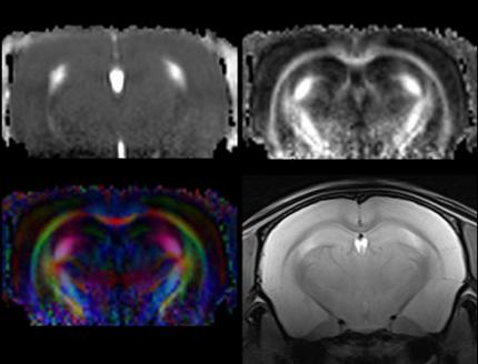
Diffusion Tensor imaging used to evaluate grey and white matter in rodent brain. Upper left – Mean Diffusivity. Upper right – Fractional Anisotropy (FA). Lower left – directionally color coded FA. Lower right – T2 weighted.
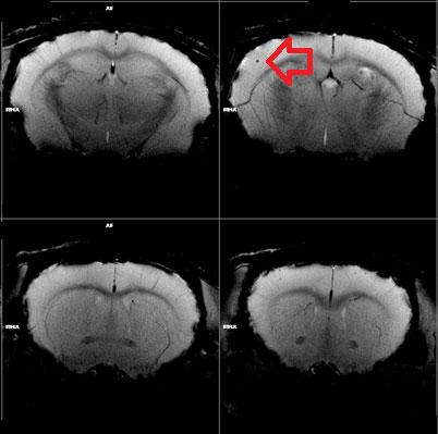
T2* images acquired with a cryocoil to detect stroke in a mouse brain.
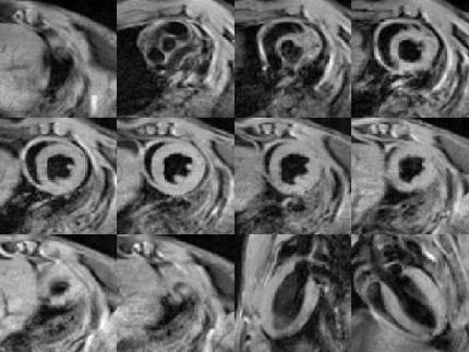
Black blood images of a beating mouse heart acquired with the mouse heart 2x2 array coil.
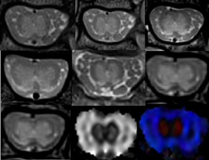
Variety of proton density, T2, fractional anisotropy (FA) map, and colored FA map images of a rat spine acquired using a 2x2 receiver array.