Pathology Research Core
What is the Pathology Research Core?
The Pathology Research core facilitates the COCVD projects by providing equipment and technical expertise to embed and section tissue specimens and perform chemical staining of tissue sections to be examined, photographed, and analyzed chromogenically by light microscopy.
Current facilities include:
- Microm STP-120 programmable tissue processor
- Leica EG1160 paraffin embedding center
- Microm HM355S microtome
- Nikon 55i Cool LED upright microscope with DS-Ri1 12MP color camera system
- Nikon 80i upright microscope with DS-Ri1 motorized stage and fluorescent capability
- Nikon Eclipse TE2000-U inverted microscope with color camera system and NIS-Elements quantitative analysis software package
Marsha Ensor, M.S. oversees the pathology research core. The previous director, Wendy Katz, developed protocols for standard tissue stains and created forms for utilization of this core.
Acknowledging the Core
Please remember when submitting publications to use the following or similar text to acknowledge the core facility: This work was supported by the UK Pathology Research core facility. (RRID:SCR_018824)

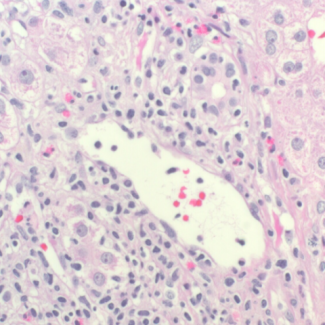
Liver biopsy slide
Pathology Research Core

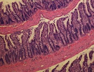
Rat intestinal swiss roll
Gipson-Reichart & Corley, H&E