Two hours of sleep restored: UK researchers make Alzheimer’s breakthrough
There’s a small fire isolated in your kitchen. If you had the right tool, you might be able to put it out. But before you can, the sprinklers turn on and flood your entire house. An automatic response to an issue has now damaged everything you own.
That’s akin to what happens in the brains of people with Alzheimer’s: amyloid plaques, sticky protein clumps that build up in the brain, are the fire in the kitchen. Microglia, the brain’s resident immune cells, are the sprinklers. A mechanism designed to protect the body ends up hurting it.
Researchers at the University of Kentucky have discovered this harmful process for the first time — and figured out how to turn it off.
In a study published in the journal Alzheimer’s & Dementia, a team led by Shannon L. Macauley, Ph.D., an associate professor of physiology in the UK College of Medicine, and first author Nicholas J. Constantino, Ph.D., a recent UK doctoral graduate, revealed that microglia are the primary drivers of sleep loss. By using a drug to temporarily remove these cells in animal models, the researchers successfully restored over two hours of sleep per day, providing a new target for treating the disease that Macauley described as “paradigm shifting.”
Previously, scientists believed that sleep loss in Alzheimer’s was caused by dying neurons or the physical clutter of amyloid plaques. However, the UK team’s findings suggest the problem is a “whole house response” scenario.
“Basically, we showed that it is not the plaques themselves, or solely dysfunctional neurons, that cause sleep loss but actually microglia,” said Macauley. “Microglia are immune cells that, when they respond to plaques, kick off this elaborate cascade of inflammation, as if the microglia are partying all night, and keeping the brain awake.”
Seeing inside
To isolate the effects of Alzheimer’s from the natural changes of aging, the team compared two groups of mice: those with a genetic predisposition to develop amyloid plaques and “wild-type” mice that aged normally. They studied these mice at six months, when plaques first emerge, and at 18 months, representing late-stage disease.
The researchers employed several sophisticated technologies to track these changes. Mice were fitted with tiny headmounts to record electroencephalography (EEG) and electromyography (EMG). An EEG acts like an electrical fingerprint of the brain by recording the unique patterns of electrical activity and oscillations produced by the brain’s networks. An EMG tracks muscle movement. This allowed the team to monitor exactly when the mice were in deep, restorative sleep, dreaming sleep or awake.
To see where the immune cells were “partying all night,” as Macauley put it, the team used a cutting-edge technique called light-sheet microscopy. This process involves making the brain tissue transparent and using a thin sheet of laser light to create a high-resolution 3D digital map. This allowed the researchers to see the entire landscape of plaques and immune cells at once.
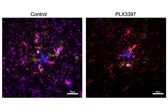
To prove that microglia were the cause of sleep loss, the team used a drug called Pexidartinib (PLX3397). This medication, originally used in cancer research, works by blocking a signal that microglia need to survive. By feeding the mice this drug for 14 days, they were able to temporarily remove 87% of the brain’s immune cells to see if sleep would return.
The team used a mathematical algorithm called Fitting Oscillations and One Over Frequency to separate the brain’s electrical activity into different parts: periodic activity (the rhythmic waves we usually think of as brain waves) and aperiodic activity (the background electrical noise).
To use a car metaphor, they were checking to see if the brain’s engine was revving too high, even during rest.
A new view
Macauley described the findings as “mind-blowing and unexpected.” The research revealed that the relationship between plaque buildup and sleep loss is not a simple, downward slide.
“I expected that as plaque burden became more severe, sleep disruption would also worsen,” said Constantino. “The disruptions in sleep and cortical EEG activity that occur at six months, when plaques first emerge, did not worsen by 18 months, despite more than double the amount of plaque burden.”
The researchers described this as a ceiling effect: even when plaque levels more than doubled, the sleep deficit remained stable. This suggests that the initial immune storm triggered by early plaques is what causes the damage, regardless of how many plaques follow.
The study also clarified how the brain changes during normal aging compared to disease. Normal aging selectively reduces rapid eye movement (REM) sleep — the stage where we dream and consolidate memories. Amyloid pathology present in Alzheimer’s selectively targets nonrapid eye movement (NREM) sleep or restorative sleep.
“That restorative sleep is super important for physical repair, learning and memory and washing out the toxins of the day,” Macauley said. “When Alzheimer’s patients lose this stage, they lose their brain’s primary cleaning cycle, creating a feed-forward loop that may drive further damage.”
2 hours of sleep restored per night
The most impactful finding was the result of the microglial depletion. When the immune cells were removed, the mice with Alzheimer’s gained more than two hours of sleep per night. Their restorative NREM sleep bouts became longer, allowing them more opportunities to transition into healthy dreaming sleep important for making new memories.
Crucially, this sleep was restored without changing the amount of amyloid plaques in the brain. This suggests that the body’s inflammatory response is a reversible cause of sleep loss that can be treated independently of the plaques themselves.
And it raises the question: can this vital sleep be restored for humans, allowing their brains to break the feed-forward loop?
The lab where it happened
This breakthrough was made possible by the unique culture of the Macauley lab in the Department of Physiology at the Sanders-Brown Center on Aging. Macauley credits the discovery to a “beautiful partnership” with her students and trainees.
“I love people who take initiative, find their passion are curious and keep pushing to find an answer,” Macauley said. She encourages her team to be “calculated risk-takers,” and keeps a quote in her office by Wayne Gretzky: “You miss 100% of the shots you don’t take.”
Lead author Constantino, who recently defended his doctorate at UK, said that this environment empowered him to ask complex, interdisciplinary questions.
“Dr. Macauley has also taught me to embrace uncertainty and failure as part of the scientific process,” Constantino said. “Some of the most interesting studies I have been a part of emerged because our original hypothesis was wrong.”
Instead of stopping at an obstacle, Macauley tells her team: “Follow the data, ask better questions and figure out what is actually happening.”
This collaborative spirit allowed the team to explore targeting microglia, rather than sticking to the traditional, neuron-centric views of the field.
What’s next?
The ultimate goal of this research is to create affordable, noninvasive tools for people facing Alzheimer’s disease, and this study accomplished several things that can be built upon in future research.
The team identified specific electrical signatures in the brain that distinguish Alzheimer’s from normal aging, and they believe EEG could be used as a “readily accessible, affordable and longitudinal biomarker of Alzheimer’s disease.”
“Portable EEG systems could allow us to monitor people in their home environments and potentially screen for changes associated with an Alzheimer’s disease, without the initial need for expensive or invasive tests,” Macauley said. This could allow doctors at local clinics across Kentucky to potentially screen those at risk before requiring individuals to travel hours for more specialized tests at major hospitals.
Macauley’s lab is now investigating how to calm down these immune cells without removing them entirely. They are exploring the use of safe, existing medications — such as the diabetes drug Metformin or antiseizure drug Stiripentol — to reset the way microglia use fuel and change how overactive these cells become. By stopping the immune cells from revving the brain’s engine, they hope to restore sleep and improve quality of life years before memory loss begins.
“If we can target that process, it might help with quality of life, attention, cognition and confusion,” Macauley said.
Solving problems starts with identifying the cause and the right tool, and Macauley’s lab is making breakthroughs in finding them both.
Research reported in this publication was supported by the National Institute on Aging of the National Institutes of Health under Award Numbers R01AG068330, R01AG093847 and P30AG072946, and by the National Institute of General Medical Sciences of the National Institutes of Health under Award Numbers P30GM127211 and P20GM148326. The content is solely the responsibility of the authors and does not necessarily represent the official views of the National Institutes of Health.
This work was supported by a $287,236 award from the Cure Alzheimer’s Fund.
This work was supported by a $250,000 award from The CART Fund (Coins for Alzheimer’s Research Trust).